Compact Bone Diagram - Copyright C The Mcgraw Hill Companies Chapter 8 Bone Bone Introduction As The Main Constituent Of The Adult Skeleton Bone Tissue Supports Fleshy Structures Protects Vital Organs Such As Those In The Cranial And Thoracic Cavities And Harbors The Bone : Add to favorites 25 favs.
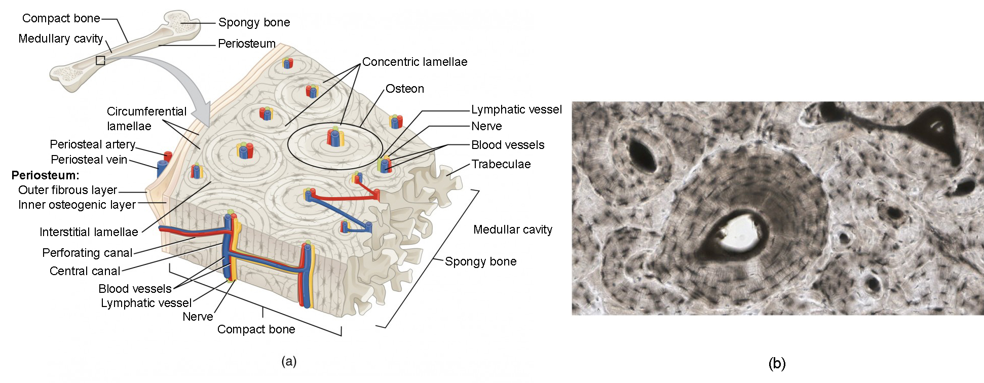
Compact Bone Diagram - Copyright C The Mcgraw Hill Companies Chapter 8 Bone Bone Introduction As The Main Constituent Of The Adult Skeleton Bone Tissue Supports Fleshy Structures Protects Vital Organs Such As Those In The Cranial And Thoracic Cavities And Harbors The Bone : Add to favorites 25 favs.. Anatomy of rib cage 12 photos of the anatomy of rib cage anatomical rib cage necklace, anatomy and physiology of rib cage, anatomy of human rib cage, anatomy of rib cage area, human anatomy rib cage muscles, human anatomy, anatomical rib cage necklace, anatomy and physiology of rib cage, anatomy of human rib cage, anatomy … (b) in this micrograph of the osteon, you can clearly see the concentric lamellae and central canals. Some, mostly older, compact bone is remodelled to form these haversian systems (or osteons). Add to favorites 0 favs. They allow blood vessels and nerves to travel through them to supply the osteocytes
The functional units of compact bone are osteons; Compact bone, also called cortical bone, is the hard, stiff, smooth, thin, white bone tissue that surrounds all bones in the human body. Compact bone, as opposed to spongy bone, is made of cylindrical units, called osteons, that are tightly formed together. The two main structural components typically include spongy bone on the interior, with an outer layer of compact bone. They allow blood vessels and nerves to travel through them to supply the osteocytes
Bone Structure Anatomy And Physiology I from s3-us-west-2.amazonaws.com It is dense (because of calcified matrix) with tiny spaces known as lucanas. Anatomy of gibt es bei ebay! Learn vocabulary, terms, and more with flashcards, games, and other study tools. Start studying compact bone labeling. Add to playlist 24 playlists. Anatomy of rib cage 12 photos of the anatomy of rib cage anatomical rib cage necklace, anatomy and physiology of rib cage, anatomy of human rib cage, anatomy of rib cage area, human anatomy rib cage muscles, human anatomy, anatomical rib cage necklace, anatomy and physiology of rib cage, anatomy of human rib cage, anatomy … (b) in this micrograph of the osteon, you can clearly see the concentric lamellae and central canals. The cells of compact bone, which is also called cortical bone, appear to be tightly packed into a solid mass.
It is dense (because of calcified matrix) with tiny spaces known as lucanas.
Terms in this set (8) spongy bone (contains red marrow) compact bone (has osteons) osteon. Add to favorites 0 favs. Compact bone, also called cortical bone, is the hard, stiff, smooth, thin, white bone tissue that surrounds all bones in the human body. Many tiny cells called osteocytes live in small spaces in the matrix and help to maintain the strength and integrity of the compact bone. Some, mostly older, compact bone is remodelled to form these haversian systems (or osteons). As seen in the image below, compact bone forms the cortex, or hard outer shell of most bones in the body. Found in short bones, flat bones, irregular bones, and end of long bones osteons cylindrical structures that comprise compact bone, organized along lines of stress, constantly changing Compact bone stands in stark contrast to trabecular bone in several ways. You need to get 100% to score the 15 points available. (b) in this micrograph of the osteon, you can clearly see the concentric lamellae and central canals. About press copyright contact us creators advertise developers terms privacy policy & safety how youtube works test new features press copyright contact us creators. Compact bone, also called cortical bone, dense bone in which the bony matrix is solidly filled with organic ground substance and inorganic salts, leaving only tiny spaces (lacunae) that contain the osteocytes, or bone cells.compact bone makes up 80 percent of the human skeleton; (b) in this micrograph of the osteon, you can clearly see the concentric lamellae and central canals.
It makes up the outer cortex of all bones and is in immediate contact with the periosteum. Compact bone, also called cortical bone, dense bone in which the bony matrix is solidly filled with organic ground substance and inorganic salts, leaving only tiny spaces (lacunae) that contain the osteocytes, or bone cells.compact bone makes up 80 percent of the human skeleton; The two main structural components typically include spongy bone on the interior, with an outer layer of compact bone. The remainder of the bone is formed by cancellous or spongy bone. Compact bone is the denser, stronger of the two types of osseous tissue (figure 6.3.6).
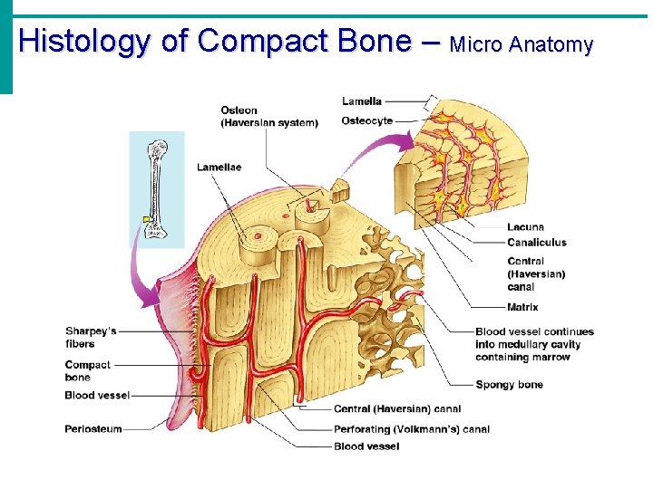
Holes Essentials Of Human Anatomy Physiology Unit 3 from slidetodoc.com Learn vocabulary, terms, and more with flashcards, games, and other study tools. Add to playlist 24 playlists. You need to get 100% to score the 15 points available. Haversian canals (sometimes canals of havers) are a series of microscopic tubes in the outermost region of bone called cortical bone. Compact bone is formed from a number of osteons, which are circular units of bone material and blood vessels. Osteocytes can be observed in the lacunae between the osteons. Online quiz to learn structure of compact bone; Deep to the compact bone layer is a region of spongy bone where the bone tissue grows in thin columns called.
Start studying compact bone labeling.
Anatomy of gibt es bei ebay! Compact bone, also called cortical bone, is the hard, stiff, smooth, thin, white bone tissue that surrounds all bones in the human body. Compact bone, as opposed to spongy bone, is made of cylindrical units, called osteons, that are tightly formed together. Related posts of compact bone diagram labeled anatomy of rib cage. Because of its strength, the compact bone makes it possible for the bone to support weight. About press copyright contact us creators advertise developers terms privacy policy & safety how youtube works test new features press copyright contact us creators. Important for compression, especially at joints. Anatomy of rib cage 12 photos of the anatomy of rib cage anatomical rib cage necklace, anatomy and physiology of rib cage, anatomy of human rib cage, anatomy of rib cage area, human anatomy rib cage muscles, human anatomy, anatomical rib cage necklace, anatomy and physiology of rib cage, anatomy of human rib cage, anatomy … As compact bone grows, osteons begin to fuse together. The remainder is cancellous bone, which has a spongelike appearance with numerous large spaces and is found in the. About press copyright contact us creators advertise developers terms privacy policy & safety how youtube works test new features press copyright contact us creators. Found in short bones, flat bones, irregular bones, and end of long bones osteons cylindrical structures that comprise compact bone, organized along lines of stress, constantly changing Serves as protection of bone marrow.
(b) in this micrograph of the osteon, you can clearly see the concentric lamellae and central canals. The remainder of the bone is formed by cancellous or spongy bone. Serves as protection of bone marrow. As seen in the image below, compact bone forms the cortex, or hard outer shell of most bones in the body. Related posts of compact bone diagram labeled anatomy of rib cage.
Draw The Given Diagram And Label The Following Parts A Spongy Boneb Periosteumc Yellow Marrowd Compact Bone from haygot.s3.amazonaws.com The remainder of the bone is formed by cancellous or spongy bone. There are two types of bone tissue: Because of its strength, the compact bone makes it possible for the bone to support weight. (b) in this micrograph of the osteon, you can clearly see the concentric lamellae and central canals. Anatomy of rib cage 12 photos of the anatomy of rib cage anatomical rib cage necklace, anatomy and physiology of rib cage, anatomy of human rib cage, anatomy of rib cage area, human anatomy rib cage muscles, human anatomy, anatomical rib cage necklace, anatomy and physiology of rib cage, anatomy of human rib cage, anatomy … The functional units of compact bone are osteons; Osteocytes can be observed in the lacunae between the osteons. In long bones, as you move from the outer cortical compact bone to the inner medullary cavity, the bone transitions to spongy bone.
(b) in this micrograph of the osteon, you can clearly see the concentric lamellae and central canals.
You need to get 100% to score the 10 points available. Compact and spongy.the names imply that the two types differ in density, or how tightly the tissue is packed together. Serves as protection of bone marrow. Deep to the compact bone layer is a region of spongy bone where the bone tissue grows in thin columns called. Some, mostly older, compact bone is remodelled to form these haversian systems (or osteons). It makes up the outer cortex of all bones and is in immediate contact with the periosteum. The diagram above shows a longitudinal view of an osteon. Terms in this set (8) spongy bone (contains red marrow) compact bone (has osteons) osteon. Anatomy of gibt es bei ebay! Haversian canals (sometimes canals of havers) are a series of microscopic tubes in the outermost region of bone called cortical bone. The remainder is cancellous bone, which has a spongelike appearance with numerous large spaces and is found in the. Which contain a centrally located haversian canal, encased in lamellae (concentric rings). You need to get 100% to score the 15 points available.


0 Comments